
Peptide Fibrils and Aggregation: When Peptides Form Unwanted Structures
Why research peptides aggregate into fibrils, amyloid-like structures, and gels. Causes, detection methods, and prevention strategies for laboratory handling.
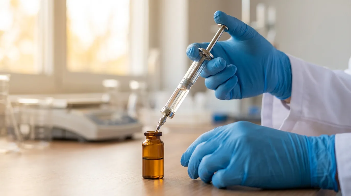
For laboratory research use only. Not for human consumption.
What Are Peptide Fibrils and Aggregates?
Peptide aggregation is the process by which individual peptide molecules associate into ordered or disordered multimeric assemblies. Fibrils represent a specific subset of aggregates characterized by elongated, unbranched structures with cross-beta-sheet architecture, where beta-strands run perpendicular to the fibril axis. These structures share morphological and structural features with amyloid fibrils observed in neurodegenerative disease research, though any peptide of sufficient length (generally 6+ residues) can potentially form fibrillar assemblies under appropriate conditions. For research laboratories working with synthetic peptides, unwanted aggregation represents a significant source of experimental variability and can compromise assay results.
Aggregates exist on a spectrum from small soluble oligomers (2-50 molecules) through protofibrils (curvilinear structures 4-10 nm in diameter) to mature fibrils (straight, rigid structures 7-13 nm wide and micrometers in length). Amorphous aggregates lack the ordered beta-sheet structure of fibrils and appear as irregular clumps under electron microscopy. Understanding which type of aggregate has formed is critical for troubleshooting peptide solubility issues in the laboratory.
Molecular Mechanisms of Peptide Aggregation
The nucleation-dependent polymerization model describes fibril formation as a two-phase process. During the lag phase, monomers undergo conformational change and associate into small oligomeric nuclei – this is thermodynamically unfavorable and rate-limiting. Once a critical nucleus forms, the elongation phase proceeds rapidly as monomers add to fibril ends in a first-order kinetic process. The sigmoidal kinetic profile (lag phase followed by exponential growth and plateau) is diagnostic for nucleated polymerization and can be monitored using Thioflavin T (ThT) fluorescence assays.
Seeding – the addition of preformed fibril fragments to fresh monomer solution – eliminates the lag phase by providing pre-formed nucleation templates. This has practical implications: contamination of peptide stocks with even trace amounts of aggregated material can accelerate aggregation of freshly reconstituted solutions. Hydrophobic interactions between non-polar side chains (Leu, Ile, Val, Phe) provide the primary driving force, supplemented by backbone hydrogen bonding that stabilizes the cross-beta architecture.
Environmental Factors That Promote Fibril Formation
Concentration is the single most important variable – aggregation propensity increases with the square of peptide concentration above the critical aggregation concentration (CAC). For most research peptides, maintaining working concentrations below 1 mg/mL significantly reduces aggregation risk. Temperature elevation accelerates aggregation kinetics by increasing molecular mobility and the frequency of productive intermolecular contacts. pH affects aggregation through its influence on net charge: peptides near their isoelectric point (pI) have minimal electrostatic repulsion and aggregate most readily.
Ionic strength modulates aggregation by screening electrostatic repulsions between charged peptide molecules. Physiological salt concentrations (150 mM NaCl) can promote aggregation of charged peptides by reducing the electrostatic energy barrier. Agitation – including vortexing, sonication, and repeated pipetting – introduces air-water interfaces that denature peptides and promote nucleation. Freeze-thaw cycles create transient high-concentration zones at ice crystal boundaries that serve as nucleation hotspots.
Peptides near their isoelectric point are most prone to aggregation. Adjusting solution pH 2-3 units away from the pI can dramatically improve solubility and reduce fibril formation.
Sequence Motifs Prone to Aggregation
Computational tools such as TANGO, Waltz, and AmylPred2 predict aggregation-prone regions (APRs) based on sequence features. Key risk factors include stretches of 5+ hydrophobic residues (VLIAF), alternating polar-nonpolar patterns that favor beta-strand formation, and aromatic residues (Phe, Tyr, Trp) that stabilize aggregates through pi-pi stacking interactions. Proline residues are strong aggregation breakers due to their inability to participate in regular beta-sheet hydrogen bonding, while charged residues (Lys, Arg, Glu, Asp) at aggregate-prone positions introduce electrostatic repulsion.
The dipeptide motif Phe-Phe (diphenylalanine) is the shortest known sequence capable of forming well-ordered nanotubes and fibrils, demonstrating the powerful role of aromatic interactions in peptide self-assembly. N-methylation of backbone amides within APRs is an effective design strategy for preventing aggregation in synthetic peptides intended for extended storage.
Analytical Methods for Detecting Aggregation
Thioflavin T (ThT) fluorescence is the most widely used screening assay for amyloid-like fibrils. ThT binds to cross-beta-sheet structures and exhibits a characteristic fluorescence enhancement (excitation 440 nm, emission 480 nm) proportional to fibril mass. Dynamic light scattering (DLS) measures hydrodynamic radius distributions and can detect oligomeric intermediates as small as 5-10 nm before visible aggregation occurs. Circular dichroism (CD) spectroscopy monitors secondary structure transitions – a shift from random coil (minimum near 198 nm) to beta-sheet (minimum near 218 nm, maximum near 195 nm) indicates structural conversion.
Transmission electron microscopy (TEM) with negative staining provides direct visualization of fibril morphology, dimensions, and periodicity. Size-exclusion chromatography (SEC) separates monomers from oligomers and higher-order aggregates based on hydrodynamic volume, and can be coupled with multi-angle light scattering (SEC-MALS) for absolute molecular weight determination of aggregate species.
Strategies to Prevent Unwanted Aggregation
Solvent selection is critical: 1,1,1,3,3,3-hexafluoro-2-propanol (HFIP) pretreatment disrupts pre-existing aggregates by stabilizing alpha-helical conformations. The standard disaggregation protocol involves dissolving the lyophilized peptide in neat HFIP at 1 mg/mL, incubating for 1 hour at room temperature, then evaporating the HFIP under nitrogen flow and reconstituting immediately in the desired buffer. This produces monomeric starting material for kinetic experiments.
Co-solvents such as DMSO (up to 5% v/v) improve solubility of hydrophobic peptides without significantly affecting most biological assays. For peptides with high aggregation propensity, working at concentrations below the CAC, maintaining pH away from pI, avoiding unnecessary agitation, and aliquoting into single-use volumes to avoid freeze-thaw cycles are effective strategies. Adding 0.01-0.05% polysorbate-20 or polysorbate-80 can prevent surface-induced aggregation at air-water and container-wall interfaces.
Storage and Handling Best Practices
Lyophilized peptides should be stored desiccated at -20C or below, with the container equilibrated to room temperature before opening to prevent moisture condensation. Reconstituted peptides are best stored as single-use aliquots at -80C in low-bind polypropylene tubes. Avoid repeated freeze-thaw cycles – each cycle increases aggregate content by approximately 2-8% depending on the peptide sequence. Polypropylene containers are preferred over glass, as glass surfaces promote adsorption and nucleation of hydrophobic peptides.
Always centrifuge reconstituted peptide solutions (16,000g, 10 min) before use to pellet any insoluble aggregates. Use only the clear supernatant for experiments.
References
- Knowles TPJ et al. (2014). The amyloid state and its association with protein misfolding diseases. Nat Rev Mol Cell Biol, 15(6):384-396.
- Hamley IW (2012). The amyloid beta peptide: a chemist's perspective. Chem Rev, 112(10):5147-5192.
- Zapadka KL et al. (2017). Factors affecting the physical stability of peptide therapeutics. Interface Focus, 7(6):20170030.
- Reches M, Gazit E (2003). Casting metal nanowires within discrete self-assembled peptide nanotubes. Science, 300(5619):625-627.
- Arosio P et al. (2015). On the lag phase in amyloid fibril formation. Phys Chem Chem Phys, 17(12):7606-7618.
- Mahler HC et al. (2009). Protein aggregation: pathways, induction factors and analysis. J Pharm Sci, 98(9):2909-2934.
- Biancalana M, Koide S (2010). Molecular mechanism of Thioflavin-T binding to amyloid fibrils. Biochim Biophys Acta, 1804(7):1405-1412.
Further Reading on ChemVerify
- Read more: How to Reconstitute Research Peptides Properly → https://www.chemverify.com/learn/how-to-reconstitute-research-peptides
- Read more: Peptide Storage Guide: Lyophilized vs. Reconstituted → https://www.chemverify.com/learn/peptide-storage-guide-lyophilized-reconstituted
References
Compare Verified Vendors
Browse COA-verified suppliers with exclusive discount codes and transparent pricing.