
Peptide Half-Life Comparison Table: From Minutes to Days
Compare the in vitro and in vivo half-lives of common research peptides. Factors affecting peptide degradation rates, protease susceptibility, and stabilization methods.
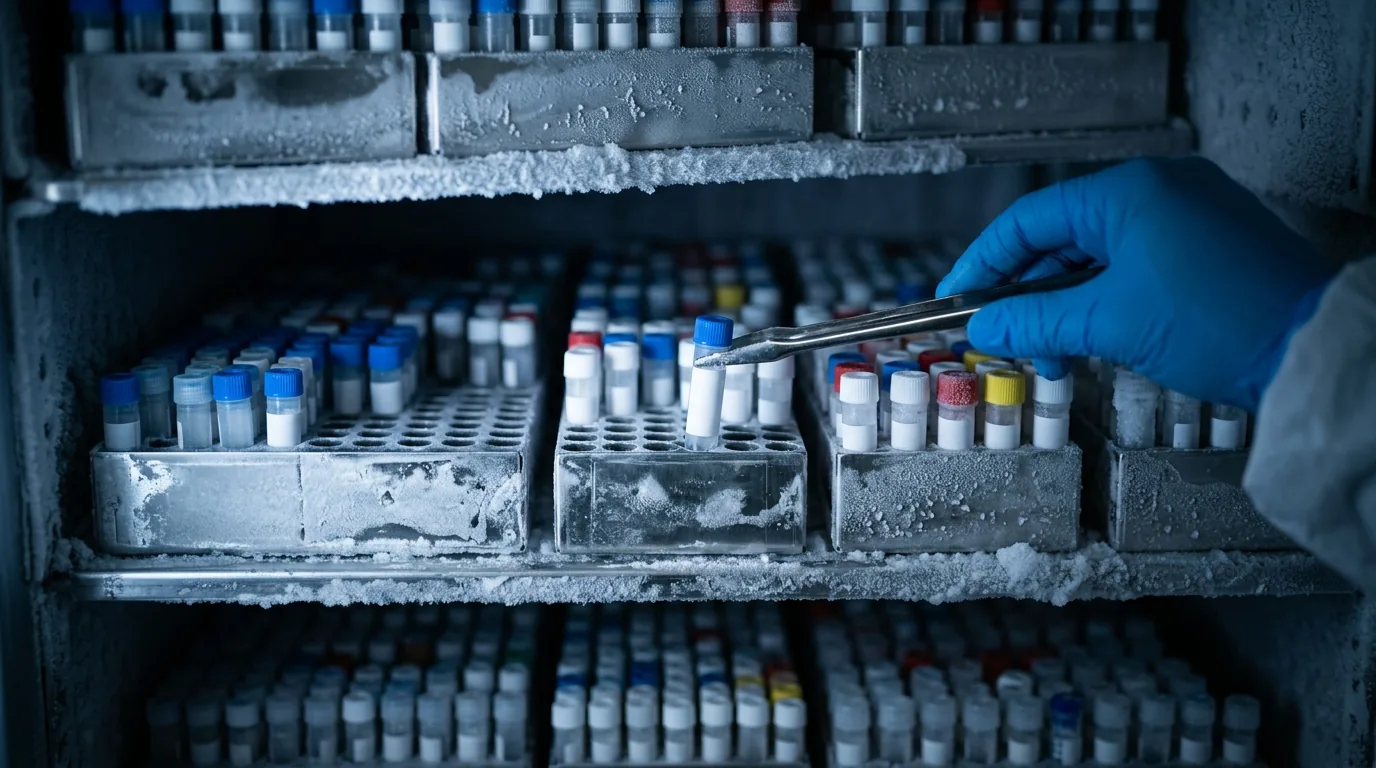
For laboratory research use only. Not for human consumption.
What Is Peptide Half-Life?
Peptide half-life (t1/2) is the time required for the concentration or biological activity of a peptide to decrease to 50% of its initial value. In research contexts, half-life can refer to several distinct measurements: chemical half-life (stability in solution at a given pH and temperature), plasma half-life (resistance to degradation by blood proteases), and elimination half-life (clearance from the body in in vivo models). These values can differ by orders of magnitude for the same peptide – a peptide chemically stable for weeks in buffered solution may be degraded within minutes in plasma.
Understanding half-life is critical for experimental design. Short-lived peptides require more frequent dosing intervals in in vivo studies, may need continuous infusion protocols, or benefit from stabilization strategies such as cyclization, D-amino acid substitution, or PEGylation. Long-lived peptides allow simpler dosing schedules but may accumulate with repeated administration.
In Vitro Half-Life: Stability in Solution and Plasma
Chemical stability in aqueous solution depends primarily on pH, temperature, and sequence-specific degradation pathways. Most linear peptides are chemically stable (t1/2 > 30 days) at pH 5-7 and 4C in the absence of proteases. The primary chemical degradation pathways – deamidation, hydrolysis, and oxidation – follow Arrhenius kinetics with typical activation energies of 80-100 kJ/mol, meaning storage at -20C versus 4C extends chemical half-life approximately 10-fold.
Plasma half-life measures resistance to enzymatic degradation and is the more pharmacologically relevant parameter. Human plasma contains a complex mixture of proteases – aminopeptidases, carboxypeptidases, endopeptidases, and dipeptidyl peptidases – that rapidly degrade most unmodified linear peptides. Plasma stability is assessed by incubating the peptide in fresh plasma (typically 25% or 100% plasma) at 37C and quantifying remaining intact peptide by LC-MS/MS at serial time points.
Half-Life Comparison of Common Research Peptides
Research peptides span a remarkable range of plasma half-lives. Very short-lived peptides (t1/2 < 5 minutes) include native GnRH (gonadotropin-releasing hormone, approximately 2-4 minutes) and bradykinin (approximately 15-30 seconds in plasma). Short-lived peptides (5-30 minutes) include native GHRH (growth hormone-releasing hormone, approximately 7-12 minutes), CJC-1295 without DAC (approximately 30 minutes), and AOD-9604 (approximately 15-25 minutes). Angiotensin II has a plasma half-life of approximately 1-2 minutes due to rapid aminopeptidase activity.
Moderate-lived peptides (30 minutes to 6 hours) include BPC-157 (estimated 60-90 minutes, limited published data), Ipamorelin (approximately 2 hours), and GHRP-6 (approximately 2-3 hours). Long-lived peptides (6+ hours) include Semaglutide (approximately 160 hours due to albumin binding via C-18 fatty acid modification), CJC-1295 with DAC (approximately 5-8 days due to covalent albumin conjugation), and Tirzepatide (approximately 5 days). These dramatic differences reflect the impact of structural modifications designed to resist proteolytic degradation and renal clearance.
Half-life values reported in literature may vary significantly depending on species (rodent vs. human plasma), concentration, assay method, and whether total or intact peptide is measured. Always note the experimental conditions when comparing published half-life data.
Factors That Determine Peptide Half-Life
Peptide length strongly influences half-life – peptides shorter than 5 residues are generally cleared within minutes by renal filtration (molecular weight cutoff approximately 5 kDa for glomerular filtration) and rapid peptidase activity. Molecular size above 40 kDa (achievable through PEGylation or fusion proteins) largely prevents renal clearance, shifting the elimination mechanism to receptor-mediated endocytosis and intracellular degradation. Charge and hydrophobicity affect plasma protein binding – highly bound peptides are shielded from both proteolysis and renal filtration, extending their apparent half-life.
Sequence composition determines protease susceptibility at specific positions. The N-terminal residue is the primary determinant of aminopeptidase susceptibility – sequences beginning with Ala, Leu, or Phe are rapidly cleaved, while N-terminal Pro, D-amino acids, or N-methylated residues resist aminopeptidases. The C-terminal residue similarly determines carboxypeptidase sensitivity. Internal cleavage sites for endopeptidases (trypsin-like enzymes cleave after Arg/Lys; chymotrypsin-like enzymes cleave after Phe/Trp/Tyr) create additional degradation points.
Key Proteases and Their Cleavage Preferences
Dipeptidyl peptidase IV (DPP-IV) cleaves dipeptides from the N-terminus of peptides containing Ala or Pro at position 2 – this enzyme is the primary reason native GLP-1 and GIP have half-lives of only 1-2 minutes. DPP-IV-resistant analogs (e.g., Semaglutide with Aib at position 2) achieve dramatically extended half-lives. Neutral endopeptidase (NEP/neprilysin) degrades many bioactive peptides including natriuretic peptides, endothelin, and substance P at hydrophobic residue sites.
Angiotensin-converting enzyme (ACE) is a dipeptidyl carboxypeptidase that cleaves C-terminal dipeptides, notably converting angiotensin I to angiotensin II and degrading bradykinin. Aminopeptidase N (APN/CD13) is a zinc metalloprotease that sequentially removes N-terminal amino acids and is particularly active in the brush border of intestinal epithelium, contributing to the poor oral bioavailability of most peptides.
Strategies to Extend Peptide Half-Life
D-amino acid substitution at protease-sensitive positions is the simplest stabilization approach – replacing L-amino acids with their D-enantiomers renders those positions invisible to most proteases. Full D-amino acid substitution (retro-inverso peptides) can produce protease-resistant analogs that retain binding affinity for certain targets. N-terminal acetylation blocks aminopeptidases, while C-terminal amidation blocks carboxypeptidases – these modifications extend half-life by 2-10 fold for many sequences with minimal impact on biological activity.
Cyclization (head-to-tail, side chain-to-side chain, or disulfide) constrains the peptide backbone and eliminates free termini, providing resistance to both exo- and endopeptidases. Stapled peptides use hydrocarbon bridges across one or two helical turns to lock alpha-helical conformations while providing protease resistance. PEGylation (attachment of polyethylene glycol chains of 20-40 kDa) dramatically increases hydrodynamic radius beyond the renal filtration cutoff and shields the peptide from protease access. Fatty acid conjugation (lipidation) enables non-covalent albumin binding in plasma, extending half-life to the timescale of albumin turnover (approximately 19 days in humans).
How Peptide Half-Life Is Measured in Research
In vitro plasma stability assays incubate the peptide (typically 1-10 uM) in 25% or 100% plasma at 37C. Aliquots are removed at predetermined time points (0, 5, 15, 30, 60, 120, 240 minutes), quenched by addition of organic solvent (acetonitrile) to precipitate proteins, and the supernatant is analyzed by LC-MS/MS for intact peptide concentration. The half-life is calculated by fitting the concentration-time data to a first-order decay equation: C(t) = C(0) * e^(-kt), where t1/2 = ln(2)/k.
In vivo pharmacokinetic studies in animal models typically administer the peptide intravenously or subcutaneously and collect serial blood samples over a time course. Plasma is separated and analyzed by LC-MS/MS or immunoassay for intact peptide. Pharmacokinetic parameters including Cmax, Tmax, AUC, clearance, and terminal half-life are derived using non-compartmental or compartmental modeling. Species differences in protease expression and renal function mean that rodent half-life data do not directly predict human values.
Practical Implications for Research Protocol Design
For in vitro cell culture experiments, short-lived peptides may require repeated media changes with fresh peptide to maintain effective concentrations over multi-day incubation periods. Adding protease inhibitor cocktails to cell culture media can extend peptide half-life in vitro but may introduce confounding effects on cell biology. Alternatively, selecting stabilized analogs (D-amino acid substituted, cyclized, or PEGylated versions) simplifies experimental protocols by reducing dosing frequency.
When designing stability studies for peptide solutions, always include a t=0 reference sample analyzed alongside later time points to control for analytical variability. Store the reference sample under conditions known to prevent degradation (e.g., -80C in single-use aliquots).
References
- Werle M, Bernkop-Schnurch A (2006). Strategies to improve plasma half life time of peptide drugs. Amino Acids, 30(4):351-367.
- Fosgerau K, Hoffmann T (2015). Peptide therapeutics: current status and future directions. Drug Discov Today, 20(1):122-128.
- Di L (2015). Strategic approaches to optimizing peptide ADME properties. AAPS J, 17(1):134-143.
- Mentlein R (1999). Dipeptidyl-peptidase IV (CD26): role in the inactivation of regulatory peptides. Regul Pept, 85(1):9-24.
- Lau JL, Dunn MK (2018). Therapeutic peptides: historical perspectives, current development trends, and future directions. Bioorg Med Chem, 26(10):2700-2707.
- Diao L, Meibohm B (2013). Pharmacokinetics and pharmacokinetic-pharmacodynamic correlations of therapeutic peptides. Clin Pharmacokinet, 52(10):855-868.
- Muttenthaler M et al. (2021). Trends in peptide drug discovery. Nat Rev Drug Discov, 20(4):309-325.
Further Reading on ChemVerify
- Read more: How Fast Do Peptides Work? Expected Timelines → https://www.chemverify.com/learn/how-fast-do-peptides-work-timelines
- Read more: Peptide Cycling: Research Duration and When to Pause → https://www.chemverify.com/learn/peptide-cycling-research-duration-pause
- Read more: What Is an Amino Acid? The Building Blocks of Peptides → https://www.chemverify.com/learn/what-is-amino-acid-building-blocks-peptides
References
Compare Verified Vendors
Browse COA-verified suppliers with exclusive discount codes and transparent pricing.