
Reconstitution Video Guide: Visual Step-by-Step for Beginners
Learn how to reconstitute lyophilized peptides safely with this visual step-by-step guide covering bacteriostatic water, sterile technique, and storage.
For laboratory research use only. Not for human consumption.
Why Proper Reconstitution Matters
Reconstitution – the process of dissolving a lyophilized (freeze-dried) peptide powder back into solution – is one of the most critical steps in peptide research. Improper reconstitution can denature the peptide, introduce contamination, produce inaccurate concentrations, or create aggregation that renders the material unusable. Understanding the correct procedure ensures that research results are reproducible and that expensive research materials are not wasted.
Lyophilized peptides are shipped as dry powders precisely because the solid state provides superior stability during transport and storage. Once reconstituted, peptides enter an aqueous environment where degradation pathways – hydrolysis, oxidation, deamidation, and aggregation – become active. This guide covers the complete procedure from solvent selection through final storage, emphasizing aseptic technique and proper documentation at each step.
Materials and Equipment Checklist
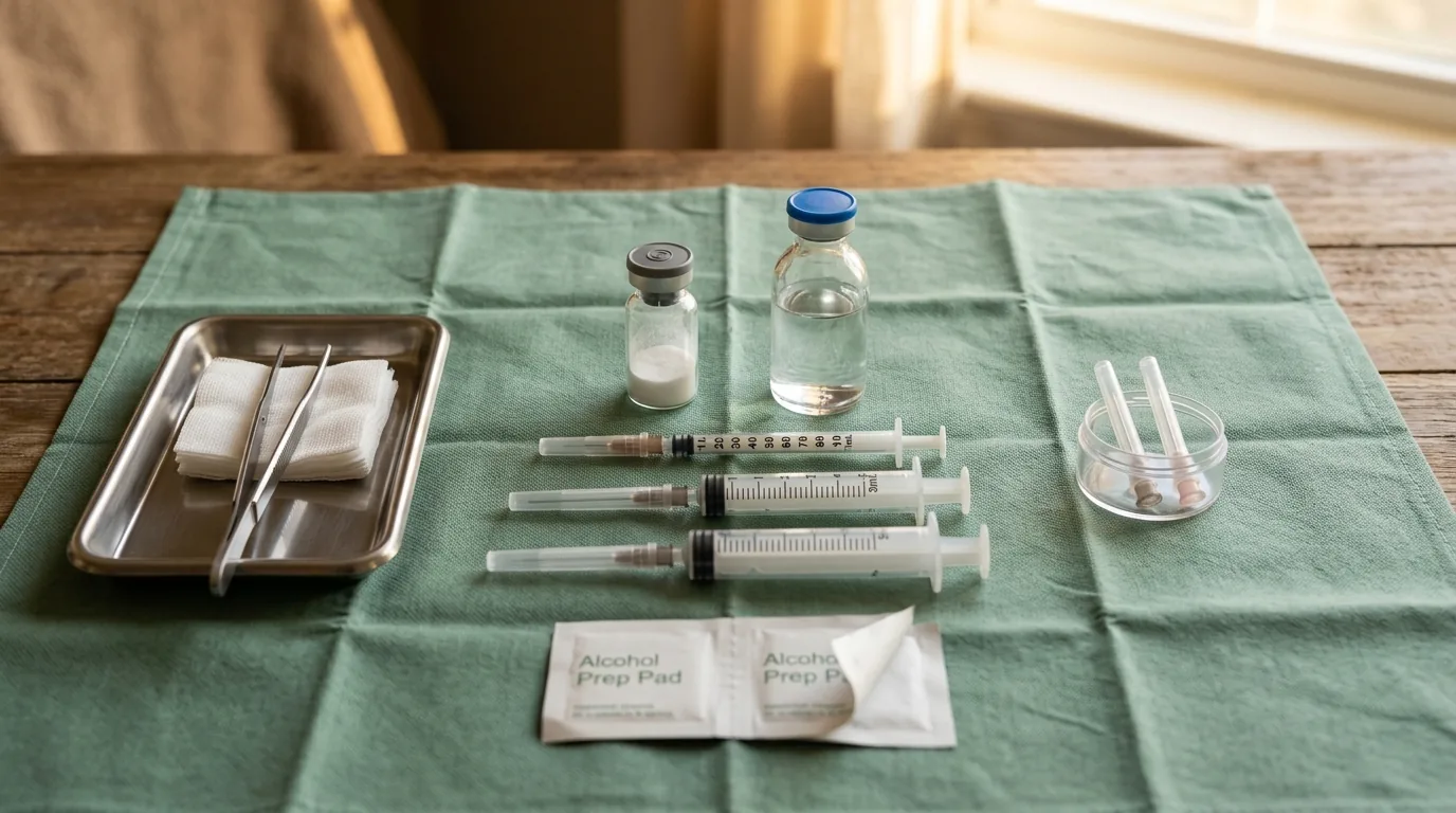
Before beginning reconstitution, gather all required materials: the lyophilized peptide vial, the appropriate solvent (bacteriostatic water, sterile water, DMSO, or dilute acetic acid depending on the peptide), alcohol swabs (70% isopropanol), appropriately sized syringes (typically 1 mL insulin syringes for volumes under 1 mL), sterile needles, and a clean workspace. Nitrile gloves should be worn throughout the procedure to prevent contamination from skin oils and microorganisms.
A laminar flow hood or biosafety cabinet provides the ideal sterile environment for reconstitution. If a laminar flow hood is unavailable, work in a clean, draft-free area that has been wiped down with 70% isopropanol. Allow the lyophilized peptide vial to reach room temperature before opening – opening a cold vial causes condensation to form inside, introducing unwanted moisture that can degrade the peptide before reconstitution even begins.
Choosing the Correct Solvent
Bacteriostatic water (sterile water containing 0.9% benzyl alcohol as a preservative) is the most commonly used reconstitution solvent for research peptides. The benzyl alcohol inhibits microbial growth, extending the usable life of the reconstituted solution. Sterile water for injection (without preservative) is appropriate when benzyl alcohol compatibility is a concern but requires single-use aliquoting and immediate freezing of unused portions.
Peptides with poor aqueous solubility may require alternative solvents. Dimethyl sulfoxide (DMSO) dissolves virtually all peptides but must be used at minimal volumes and further diluted into aqueous buffer. Dilute acetic acid (0.1% v/v) is preferred for highly basic peptides, while dilute ammonium hydroxide (0.1% v/v) improves solubility of acidic peptides. The vendor certificate of analysis (CoA) often includes a recommended reconstitution solvent – always check this document first.
Step-by-Step Reconstitution Procedure
Step 1: Clean the workspace and put on fresh nitrile gloves. Wipe the vial stopper and solvent vial with an alcohol swab and allow to air dry for 30 seconds. Step 2: Using an appropriately sized syringe, draw the calculated volume of solvent. Step 3: Insert the needle through the vial stopper at a slight angle and direct the solvent stream against the glass wall of the vial – never squirt solvent directly onto the lyophilized cake, as the mechanical force can cause foaming and denaturation.
Step 4: Allow the solvent to trickle down the vial wall onto the peptide powder. Step 5: Once all solvent has been added, gently swirl the vial in a circular motion for 30-60 seconds. Do not shake, vortex, or invert the vial vigorously – mechanical agitation generates air-liquid interfaces that promote peptide aggregation. Step 6: If undissolved material remains, let the vial sit at room temperature for 5-10 minutes, then swirl again gently. Step 7: Label the vial with the peptide name, concentration, reconstitution date, and solvent used.
Calculating Peptide Concentration
The reconstituted concentration is calculated by dividing the mass of peptide in the vial by the volume of solvent added. The mass should be taken from the vendor's certificate of analysis (actual fill weight), not the nominal label amount. For example, if a vial labeled as 5 mg actually contains 4.8 mg (per the CoA) and is reconstituted in 2.0 mL, the true concentration is 2.4 mg/mL, not 2.5 mg/mL.
For molar concentration calculations, divide the mass concentration (mg/mL) by the molecular weight (g/mol) and multiply by 1,000 to convert to micromolar. A peptide with MW 1,200 g/mol at 2.4 mg/mL has a molar concentration of 2,000 micromolar (2 mM). Recording both mass and molar concentrations on the vial label eliminates conversion errors during experimental setup.
Common Reconstitution Mistakes to Avoid
The most frequent reconstitution error is forceful injection of solvent directly onto the lyophilized cake. This creates persistent foam that traps peptide at the air-liquid interface, reducing recoverable yield by 10-30% for hydrophobic peptides. Always direct the solvent stream against the glass vial wall. The second most common error is using the wrong solvent – reconstituting a hydrophobic peptide in pure water often produces a cloudy suspension of aggregates.
Using contaminated syringes or needles introduces bacteria that can degrade the peptide within hours. Always use fresh, individually packaged sterile syringes. Repeatedly piercing the vial stopper with large-gauge needles fragments the rubber, generating particulate contamination. Use the smallest needle gauge practical for solvent addition and limit the number of punctures. Finally, failing to allow the vial to equilibrate to room temperature before opening leads to moisture condensation.
Storage After Reconstitution
Reconstituted peptide solutions should be stored at 2-8 degrees Celsius for short-term use (up to 2-4 weeks when reconstituted in bacteriostatic water) or at -20 degrees Celsius for longer-term storage. Aliquoting the reconstituted solution into single-use portions before freezing avoids repeated freeze-thaw cycles, which promote aggregation and oxidation. Use low-protein-binding microcentrifuge tubes (polypropylene) to minimize surface adsorption losses.
Reconstituted peptides in sterile water (without preservative) should be used within 24-48 hours or immediately frozen as single-use aliquots. Each freeze-thaw cycle reduces peptide recovery by an estimated 5-15% depending on the sequence. For valuable peptides, adding a cryoprotectant such as 0.1% bovine serum albumin (BSA) or 5% trehalose can reduce freeze-thaw losses significantly. Always document the number of freeze-thaw cycles on the vial label.
Troubleshooting Reconstitution Problems
If the peptide does not dissolve within 10 minutes of gentle swirling, the solvent choice may be incorrect. Add a small volume (5-10% of total) of DMSO to the vial, swirl to dissolve, then add the remaining volume of aqueous solvent. If a cloudy or turbid solution forms, the peptide may be aggregating – try brief sonication in a water bath for 30 seconds at room temperature. If turbidity persists after sonication, the peptide-solvent system is incompatible.
Gel-like material at the bottom of the vial indicates severe aggregation, typically caused by reconstitution at too high a concentration or in an incompatible solvent. This is often irreversible. If the peptide appears discolored (yellow or brown), oxidation has occurred – this is most common with methionine- or tryptophan-containing peptides exposed to light or elevated temperatures. Oxidized material should not be used for quantitative research.
References
- Manning MC et al. (2010). Stability of protein pharmaceuticals: an update. Pharm Res, 27(4):544-575.
- Chi EY et al. (2003). Physical stability of proteins in aqueous solution. Pharm Res, 20(9):1325-1336.
- Wang W (2005). Protein aggregation and its inhibition in biopharmaceutics. Int J Pharm, 289(1-2):1-30.
- Carpenter JF et al. (1997). Rational design of stable lyophilized protein formulations. Pharm Biotechnol, 10:109-133.
- Pikal MJ (2004). Mechanisms of protein stabilization during freeze-drying. Pharm Biotechnol, 14:63-107.
- Cleland JL et al. (1993). The development of stable protein formulations. Crit Rev Ther Drug Carrier Syst, 10(4):307-377.
- Bhatnagar BS et al. (2007). Protein stability during freezing. Pharm Res, 24(4):720-733.
Further Reading on ChemVerify
- Read more: Peptide Solvent Compatibility Guide → https://www.chemverify.com/learn/peptide-solvent-compatibility-guide
- Read more: Peptide Storage Temperature Guide → https://www.chemverify.com/learn/peptide-storage-temperature-guide-freeze-refrigerate
References
Compare Verified Vendors
Browse COA-verified suppliers with exclusive discount codes and transparent pricing.