
TRH (Thyrotropin-Releasing Hormone): Research Guide & Chemical Profile
TRH (pGlu-His-Pro-NH2) is a hypothalamic tripeptide releasing TSH and prolactin. MW 362.38, neuroprotective research, and neuroendocrine signaling reviewed.
For laboratory research use only. Not for human consumption.
What Is TRH (Thyrotropin-Releasing Hormone)?
TRH (thyrotropin-releasing hormone) is a hypothalamic tripeptide with the structure pyroglutamyl-histidyl-proline amide (pGlu-His-Pro-NH2) and a molecular weight of 362.38 Da. First characterized by Roger Guillemin and Andrew Schally in 1969 – work that earned them the 1977 Nobel Prize in Physiology or Medicine – TRH was the first hypothalamic releasing hormone to have its structure determined. It stimulates the release of thyroid-stimulating hormone (TSH) and prolactin from the anterior pituitary gland, but subsequent research has revealed a far broader spectrum of activity including neuroprotective, analeptic, and neuromodulatory effects that are independent of its thyroid axis function.
Tripeptide Structure: pGlu-His-Pro-NH2
TRH is one of the smallest biologically active peptide hormones. Its three residues – pyroglutamic acid (pGlu), histidine (His), and proline amide (Pro-NH2) – are both N- and C-terminally modified, which confers significant resistance to generic aminopeptidases and carboxypeptidases. The N-terminal pyroglutamate is a cyclized form of glutamic acid that blocks aminopeptidase attack, while the C-terminal amide group prevents carboxypeptidase degradation. These modifications give TRH a plasma half-life of approximately 5-6 minutes – short but notably longer than unprotected tripeptides.
The molecular formula is C16H22N6O4 with a molecular weight of 362.38 Da. TRH is freely soluble in water, slightly soluble in ethanol, and adopts a beta-turn conformation in solution as demonstrated by NMR studies. The imidazole side chain of histidine (pKa approximately 6.0) is critical for receptor binding – methylation or replacement of this residue abolishes biological activity in all assay systems tested.
Both terminal modifications of TRH (N-terminal pyroglutamate and C-terminal amide) are essential for biological activity. The unmodified linear form Glu-His-Pro shows no TRH receptor binding.
Hypothalamic-Pituitary-Thyroid Axis Role
TRH is synthesized in the paraventricular nucleus (PVN) of the hypothalamus and released into the hypophyseal portal circulation, where it reaches thyrotroph cells in the anterior pituitary. Binding to TRH receptor type 1 (TRHR1) on thyrotrophs activates phospholipase C (PLC) via Gq/11 coupling, generating inositol trisphosphate (IP3) and diacylglycerol (DAG). IP3-mediated calcium release from the endoplasmic reticulum triggers TSH exocytosis, while DAG-activated protein kinase C (PKC) sustains the secretory response.
TSH release in response to TRH is subject to negative feedback from circulating thyroid hormones (T3 and T4). T3 suppresses TRH gene expression in the PVN (via thyroid hormone receptor beta) and simultaneously reduces TRHR1 expression on thyrotrophs, creating a dual feedback mechanism. The TRH stimulation test – measuring TSH response to intravenous TRH – was historically used to diagnose secondary and tertiary hypothyroidism, though it has been largely replaced by sensitive TSH assays in clinical practice.
TRH Receptor Pharmacology (TRHR1 and TRHR2)
Two TRH receptor subtypes have been identified in mammals: TRHR1 and TRHR2. Both are G protein-coupled receptors (GPCRs) of the rhodopsin family, coupled primarily to Gq/11 proteins. TRHR1 is the dominant subtype in the anterior pituitary and mediates TSH and prolactin release. TRHR2 is expressed predominantly in the central nervous system – particularly in the thalamus, hypothalamus, cerebral cortex, and hippocampus – and is believed to mediate the extra-thyroidal neurological effects of TRH.
Both receptors bind TRH with similar affinity (Kd approximately 5-10 nM) but show differential desensitization kinetics. TRHR1 undergoes rapid agonist-induced internalization via beta-arrestin-dependent mechanisms with a t1/2 of approximately 15 minutes, while TRHR2 shows slower internalization kinetics (t1/2 approximately 45 minutes). This difference may contribute to the sustained neurological effects of TRH compared to its more transient endocrine effects.
Neuroprotective Research Applications
TRH and its analogs have demonstrated neuroprotective effects in multiple preclinical models. In traumatic brain injury (TBI) models, TRH administration reduced cerebral edema, improved blood-brain barrier integrity, and attenuated neuronal apoptosis. A 1996 study in rodent closed-head injury models showed that TRH at 10 mg/kg IV reduced contusion volume by 35% when administered within 1 hour of injury. The neuroprotective mechanism involves suppression of excitotoxic glutamate release, stabilization of neuronal membrane potential via modulation of sodium and calcium channel conductance, and attenuation of post-traumatic lipid peroxidation.
In spinal cord injury research, TRH and its stable analog CG3703 improved functional motor recovery scores in rat contusion models. The analeptic (arousal-promoting) properties of TRH have also been investigated in models of sedation and respiratory depression, where TRH antagonizes the effects of opioids, barbiturates, and ethanol on consciousness and respiration – effects that are independent of its endocrine actions and are mediated through TRHR2 in brainstem nuclei.
Prolactin Release and Neuroendocrine Effects
In addition to TSH, TRH is a potent stimulator of prolactin release from lactotroph cells in the anterior pituitary. The mechanism is identical to TSH release – Gq/11-coupled PLC activation and calcium-dependent exocytosis. TRH-stimulated prolactin release is approximately 3-5 fold in magnitude and peaks at 15-30 minutes following IV administration in human subjects. This prolactin-releasing effect has been used as a pharmacological tool to assess lactotroph function and dopaminergic tone in neuroendocrine research.
TRH neurons in the hypothalamus also project to extrahypothalamic brain regions including the nucleus accumbens, amygdala, and brainstem reticular formation. These projections mediate effects on arousal, locomotor activity, body temperature, gastric motility, and cardiovascular function that are distinct from the endocrine actions of TRH. The widespread CNS distribution of TRH and its receptors has led to its characterization as a neuromodulator with functions extending far beyond its original identification as a releasing hormone.
Metabolic Degradation and Peptidase Pathways
Despite its terminal modifications, TRH is rapidly degraded in serum and tissue by a specific serine protease known as TRH-degrading ectoenzyme (TRH-DE, also called pyroglutamyl peptidase II). This enzyme cleaves the pGlu-His bond, yielding cyclo(His-Pro) – also known as histidyl-proline diketopiperazine – and free pyroglutamic acid. Notably, cyclo(His-Pro) itself has biological activity, including effects on food intake, glucose metabolism, and neuroprotection, suggesting that TRH degradation generates a bioactive metabolite rather than simply inactivating the parent hormone.
TRH-DE expression is regulated by thyroid hormones – T3 increases TRH-DE activity in the anterior pituitary, providing an additional mechanism for negative feedback regulation. Inhibitors of TRH-DE have been developed as potential therapeutic agents to prolong TRH activity, with some showing efficacy in preclinical models of depression and cognitive impairment.
Analytical Identification and Purity
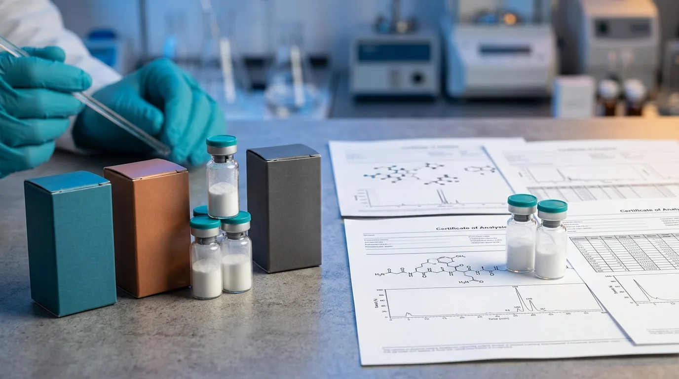
Research-grade TRH is characterized by HPLC purity of 98% or higher, confirmed by reversed-phase C18 chromatography using a water/acetonitrile gradient with 0.1% TFA. Mass spectrometry should confirm the [M+H]+ ion at m/z 363.4. The pyroglutamate modification must be verified – the linear Glu-His-Pro-NH2 precursor has a different retention time on HPLC and is biologically inactive. Optical rotation measurements can confirm the L-configuration of all three amino acids. Certificates of Analysis should include amino acid analysis, water content (Karl Fischer), and endotoxin testing.
Confirm the pyroglutamate cyclization on TRH COAs. The linear Glu-His-Pro-NH2 precursor co-elutes closely on some HPLC methods but has no TRH receptor activity.
References
- Boler J, Enzmann F, Folkers K, Bowers CY, Schally AV. (1969). The identity of chemical and hormonal properties of TRH. Biochem Biophys Res Commun, 37(4):705-710.
- Faden AI et al. (1999). TRH analogs in CNS injury. Ann NY Acad Sci, 553:380-384.
- Heuer H et al. (2000). Expression of TRH receptor subtypes in the brain. Neuroendocrinology, 72(3):151-159.
- Sun Y et al. (2008). TRH neuroprotection in spinal cord injury. J Neurotrauma, 25(7):847-856.
- Pekary AE, Sattin A. (2012). TRH in CNS aging and neurodegenerative diseases. Peptides, 38(2):322-328.
- Charli JL et al. (1998). TRH-degrading ectoenzyme. Neurochem Int, 32(4):429-436.
- Bowers CY. (2001). TRH discovery and significance. Thyroid, 11(3):271-280.
- Gary KA et al. (2003). TRH analogs: structure-activity relationships. Curr Pharm Des, 9(6):471-487.
Further Reading on ChemVerify
- Read more: B7-33: Research Guide & Chemical Profile → https://www.chemverify.com/learn/b7-33-research-guide-chemical-profile
- Read more: Ipamorelin + CJC-1295 (No DAC) Stack: Synergy Research Guide → https://www.chemverify.com/learn/ipamorelin-cjc-1295-no-dac-stack-synergy
- Read more: TP508 (Chrysalin): Research Guide & Chemical Profile → https://www.chemverify.com/learn/tp508-chrysalin-research-guide-chemical-profile
- Read more: Semax for Cognitive Research: ACTH(4-10) Analog Mechanism → https://www.chemverify.com/learn/semax-cognitive-research-acth-mechanism
References
Compare Verified Vendors
Browse COA-verified suppliers with exclusive discount codes and transparent pricing.
Continue Reading
FOXO4-DRI: Complete Research Guide & Chemical Profile
Comprehensive chemical profile of FOXO4-DRI, a D-retro-inverso peptide that disrupts the FOXO4-p53 interaction in senescent cells. Covers senolytic mechanism, cellular senescence research, and aging biology applications.
KPV: Complete Research Guide & Chemical Profile
Comprehensive chemical profile of KPV (Lys-Pro-Val), a C-terminal tripeptide fragment of alpha-MSH with MW 342.43 Da. Covers NF-kB inhibition, gut inflammation research, melanocortin signaling, and PepT1 transport.
Thymalin: Complete Research Guide & Chemical Profile
Complete research guide to Thymalin (Glu-Trp dipeptide), a thymic bioregulator peptide investigated for immune modulation, T-cell differentiation, and aging research by Khavinson and colleagues.
ARA 290 (Cibinetide): Research Guide & Chemical Profile
ARA 290 (Cibinetide) is an 11-amino-acid EPO-derived peptide targeting the innate repair receptor. Chemical profile, neuropathy and sarcoidosis research reviewed.