
Disulfide Bond Formation in Synthetic Peptides: Chemistry, Methods, and Verification
Technical guide to disulfide bond formation in research peptides – cysteine oxidation chemistry, regioselective strategies for multiple disulfide bonds, orthogonal protection schemes, and analytical verification methods.
For laboratory research use only. Not for human consumption.
TL;DR: Disulfide bonds between cysteine residues are critical for peptide tertiary structure and biological activity. Formation strategies include air oxidation, DMSO-mediated oxidation, iodine oxidation, and orthogonal protecting group schemes for regioselective multi-disulfide peptides. Verification requires mass spectrometry (free vs. oxidized mass difference of 2 Da per bond), Ellman's assay for free thiols, and HPLC comparison of reduced vs. oxidized forms.
Last verified: March 2026 | Data accuracy confirmed by ChemVerify Editorial Team
Disulfide Bond Chemistry
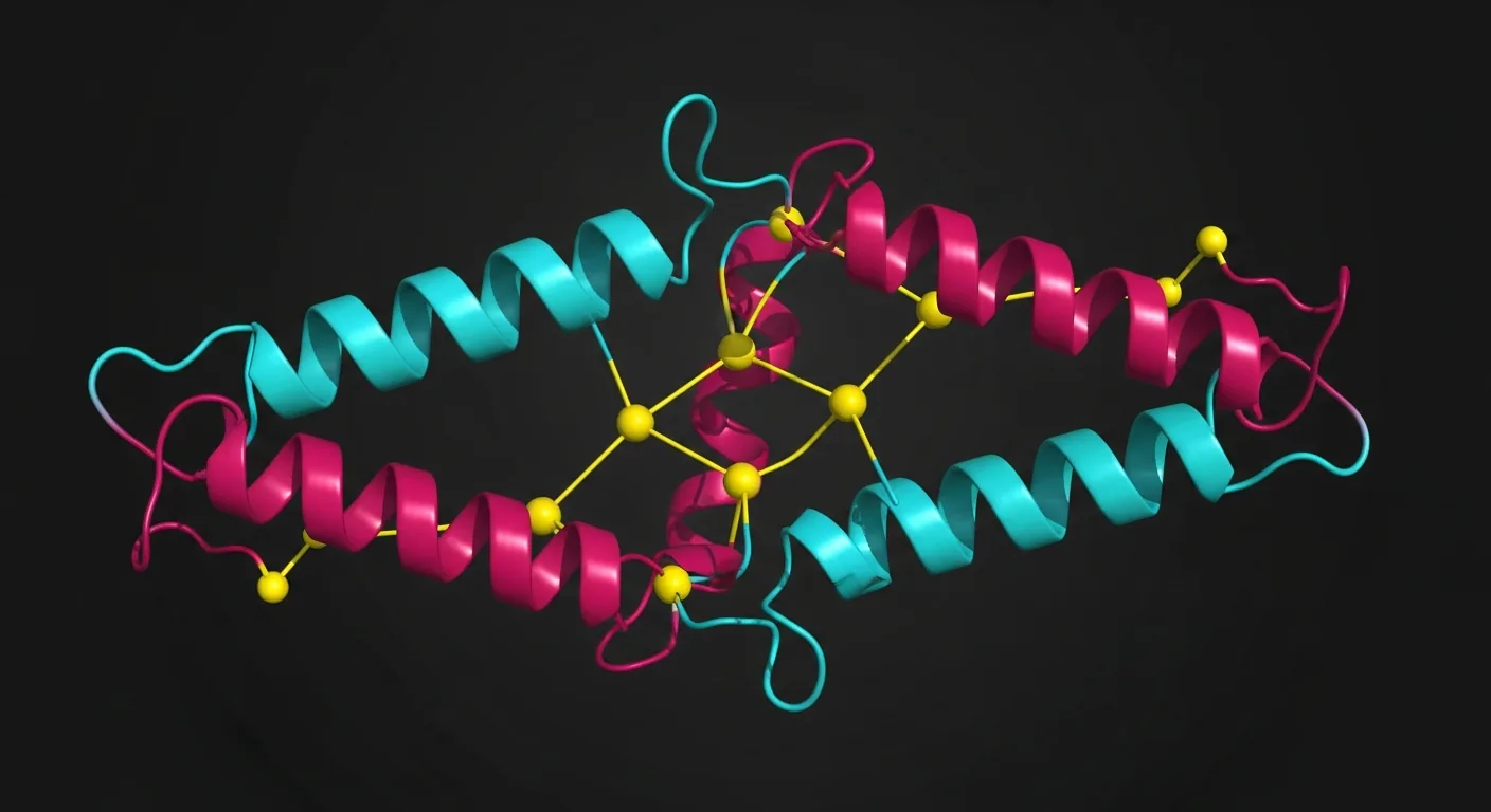
Disulfide bonds (S–S) form through the oxidation of two cysteine thiol (–SH) groups, creating a covalent cystine linkage. This reaction involves a two-electron oxidation with a standard reduction potential of approximately −0.25 V at pH 7.0. Disulfide bonds are critical structural elements in many biologically active peptides, stabilizing three-dimensional conformations and often being essential for biological activity.
The general reaction is: 2 R–SH → R–S–S–R + 2H⁺ + 2e⁻. In synthetic peptide chemistry, this oxidation must be carefully controlled to ensure the correct cysteine pairing, particularly in peptides containing more than two cysteine residues.
Single Disulfide Bond Formation
For peptides containing exactly two cysteine residues, a single intramolecular disulfide bond can be formed using several oxidation methods:
Air Oxidation
The simplest method involves dissolving the reduced peptide at low concentration (0.01–0.5 mg/mL) in a slightly basic buffer (pH 7.5–8.5, typically ammonium bicarbonate or Tris-HCl) and stirring exposed to air for 12–48 hours. Low concentration favors intramolecular cyclization over intermolecular oligomerization. Yields vary from 30–80% depending on the sequence.
DMSO Oxidation
Dimethyl sulfoxide (DMSO) at 5–20% v/v in aqueous buffer (pH 6–8) accelerates disulfide formation relative to air oxidation, typically completing in 1–6 hours. DMSO acts as a mild oxidant (E° = +0.16 V), reducing to dimethyl sulfide (DMS). This method is widely used for its simplicity and compatibility with most sequences.
Glutathione Redox Buffer
A mixture of reduced (GSH) and oxidized (GSSG) glutathione at ratios of 1:1 to 10:1 (GSH:GSSG, total concentration 1–5 mM) in buffer at pH 7.5–8.5 establishes a redox equilibrium that allows thermodynamic control of disulfide formation. This method favors the most stable (native) disulfide pairing and permits reshuffling of incorrectly formed bonds.
Iodine Oxidation
Molecular iodine (I₂) in acetic acid/methanol or aqueous solution rapidly oxidizes free thiols to disulfides. This is a fast (minutes) but harsh method that can cause side reactions with tryptophan, tyrosine, and methionine residues. Typically used at 10–50 equivalents of I₂ relative to the peptide.
Multiple Disulfide Bonds
Peptides with more than two cysteines present a combinatorial challenge. For n cysteine pairs, the number of possible disulfide isomers is (2n)! / (2ⁿ × n!). A peptide with 4 cysteines (2 disulfide bonds) has 3 possible isomers; with 6 cysteines (3 disulfide bonds), there are 15 possible isomers. Only one isomer typically represents the desired product.
Random Oxidation
Oxidative folding using air or glutathione redox buffers can yield the correct isomer if it represents the thermodynamically most stable configuration. This approach works well for peptides that fold cooperatively into a defined structure. Success rates are sequence-dependent – some peptides fold efficiently (>50% correct isomer), while others produce complex mixtures requiring extensive purification.
Orthogonal Protection Strategies
Regioselective disulfide bond formation uses orthogonal cysteine protecting groups that can be removed independently, allowing sequential, directed oxidation of specific cysteine pairs:
- Trityl (Trt): Removed by dilute TFA (1–2%) or iodine. Standard Fmoc-SPPS protecting group.
- Acetamidomethyl (Acm): Removed by iodine, mercury(II) acetate, or thallium(III) trifluoroacetate. Stable to TFA and piperidine.
- tert-Butylthio (StBu): Removed by thiol exchange (DTT, TCEP, or β-mercaptoethanol). Orthogonal to Trt and Acm.
- Methylbenzyl (MeBzl): Removed by HF or TFMSA. Used in Boc-SPPS.
- Phenylacetamidomethyl (Phacm): Removed enzymatically by penicillin G acylase. Mild, selective conditions.
A typical two-disulfide strategy: (1) Remove Trt groups during cleavage → oxidize first disulfide by air/DMSO, (2) Remove Acm groups with iodine → simultaneously forms second disulfide. This sequential approach achieves regioselectivities of 40–80% for the desired isomer, compared to 10–33% for random oxidation of 4-cysteine peptides.
Analytical Verification
Free Thiol Quantification
Ellman's reagent (5,5'-dithio-bis-(2-nitrobenzoic acid), DTNB) reacts with free thiol groups to release the yellow chromophore 2-nitro-5-thiobenzoate (TNB²⁻), measured at 412 nm (ε = 14,150 M⁻¹cm⁻¹). A fully oxidized peptide should show zero free thiols. Residual free thiol content above 5% of theoretical indicates incomplete oxidation.
Mass Spectrometry
Each disulfide bond results in a mass decrease of 2.016 Da (loss of two hydrogen atoms). For a peptide with n disulfide bonds, the expected mass shift is −(n × 2.016) Da relative to the fully reduced form. High-resolution MS can confirm the number of disulfide bonds present.
HPLC Comparison
Reduced and oxidized forms of a peptide typically exhibit different RP-HPLC retention times. The oxidized (cyclized) form is generally more compact and elutes earlier than the reduced (linear) form. Comparison of retention times before and after reduction with TCEP or DTT confirms disulfide bond presence.
Enzymatic Mapping
For multiple disulfide bonds, enzymatic digestion (trypsin, chymotrypsin) under non-reducing conditions followed by LC-MS/MS can map specific disulfide connectivities. Disulfide-linked peptide fragments are identified by their composite mass and confirmed by MS/MS fragmentation.
CoA Considerations for Disulfide Peptides
- The CoA should specify whether the peptide is supplied in reduced (linear) or oxidized (disulfide) form
- For oxidized peptides: free thiol content should be reported (acceptable: < 5% of theoretical)
- Mass spectrum should show the expected mass for the oxidized form (−2n Da from reduced)
- HPLC purity should be determined under non-reducing conditions to preserve disulfide bonds
- For multiple disulfide bonds: the specific disulfide connectivity (e.g., Cys1-Cys4, Cys2-Cys5) should ideally be verified and reported
- Peptides requiring specific disulfide pairings should include evidence of regioselectivity (enzymatic mapping or NMR data)
Frequently Asked Questions
Why are disulfide bonds important in synthetic peptides?
Disulfide bonds stabilize the three-dimensional conformation of peptides, which is essential for receptor binding and biological activity. Peptides like oxytocin, insulin, and somatostatin require correct disulfide connectivity to function. Incorrect or missing bonds produce inactive or misfolded products.
What is the difference between random and regioselective disulfide formation?
Random oxidation allows all free cysteines to pair spontaneously, which works for single-disulfide peptides but produces statistical mixtures for multi-cysteine peptides. Regioselective strategies use orthogonal protecting groups (Acm, Trt, StBu) to form specific bonds sequentially, ensuring correct connectivity in complex multi-disulfide targets.
How do I confirm a disulfide bond has formed correctly?
Use ESI-MS or MALDI-MS to confirm a mass reduction of 2 Da per disulfide bond. Ellman's reagent (DTNB) quantifies remaining free thiols. Comparative RP-HPLC shows retention time shifts between reduced and oxidized forms. For multi-disulfide peptides, enzymatic digestion with mass mapping confirms specific bond assignments.
Which oxidation method is best for single-disulfide peptides?
For single-disulfide peptides, air oxidation in dilute aqueous buffer (pH 7.5–8.5, 0.01–0.1 mg/mL) is simplest but slow (12–48 hours). DMSO (10–20% in aqueous buffer) accelerates oxidation to 1–6 hours. Iodine oxidation is fastest but can modify tryptophan and methionine residues if present in the sequence.
Can disulfide bonds form during SPPS on-resin?
On-resin disulfide formation is possible using iodine treatment of Acm-protected cysteines or thallium(III) trifluoroacetate oxidation. However, solution-phase oxidation after full cleavage and deprotection is more common and generally provides better yields for most peptide targets.
Further Reading on ChemVerify
- Read more: Acetate vs Arginate Salt Forms in Peptides: Which Is Better? → https://www.chemverify.com/learn/acetate-vs-arginate-salt-peptides-comparison
- Read more: Peptide Aggregation: Why Peptides Clump and How to Prevent It → https://www.chemverify.com/learn/peptide-aggregation-clumping-prevention
- Read more: Peptide Degradation: Deamidation, Oxidation, and How to Prevent It → https://www.chemverify.com/learn/peptide-degradation-deamidation-oxidation-prevention
- Read more: Peptide Modifications: PEGylation, Lipidation, Cyclization, and D-Amino Acids → https://www.chemverify.com/learn/peptide-modifications-pegylation-lipidation-cyclization
References
Compare Verified Vendors
Browse COA-verified suppliers with exclusive discount codes and transparent pricing.
Continue Reading
Peptide Degradation: Deamidation, Oxidation, and How to Prevent It
Scientific guide to peptide degradation pathways including asparagine deamidation, methionine oxidation, disulfide scrambling, and evidence-based prevention strategies.
Peptide Aggregation: Why Peptides Clump and How to Prevent It
Understanding peptide aggregation in research: hydrophobic clumping, amyloid formation, concentration and pH effects, co-solvents, gentle mixing, and filtration strategies.
Peptide Modifications: PEGylation, Lipidation, Cyclization, and D-Amino Acids
Advanced guide to peptide chemical modifications including PEG conjugation, fatty acid acylation, stapled peptides, head-to-tail cyclization, retro-inverso design, and non-natural amino acids.
Acetate vs Arginate Salt Forms in Peptides: Which Is Better?
A comparative analysis of acetate and arginate counterion salt forms in synthetic peptides, covering their impact on solubility, stability, pH of reconstituted solutions, bioavailability, TFA contamination concerns, and how to identify salt form on a certificate of analysis.