
Melanotan 1 vs Melanotan 2: MSH Analogs Compared
Structural comparison of Melanotan I (afamelanotide, linear 13-aa α-MSH analog, ~1,646 Da) and Melanotan II (cyclic 7-aa lactam, ~1,024 Da). Covers receptor selectivity, stability, and chromatographic profiles.
For laboratory research use only. Not for human consumption.
TL;DR: Melanotan I (afamelanotide) is a linear 13-amino-acid analog of α-MSH (~1,646 Da) with selectivity for MC1R. Melanotan II is a cyclic lactam heptapeptide (~1,024 Da) with broader melanocortin receptor activity across MC1R, MC3R, MC4R, and MC5R. The structural difference—linear vs cyclic—drives divergent receptor selectivity, metabolic stability, and analytical profiles.
Last verified: April 2026 | Data accuracy confirmed by ChemVerify Editorial Team
Introduction: Linear vs Cyclic Melanocortin Analogs
Melanotan I and Melanotan II are synthetic analogs of alpha-melanocyte-stimulating hormone (α-MSH), both developed at the University of Arizona in the 1980s-1990s. Despite sharing the melanocortin pharmacophore His-Phe-Arg-Trp, these compounds differ fundamentally in their molecular architecture: Melanotan I is a linear tridecapeptide, while Melanotan II is a cyclic heptapeptide constrained by a lactam bridge. This structural divergence produces markedly different receptor selectivity profiles, metabolic stabilities, and analytical characteristics [1][2].
For researchers in melanocortin biology, understanding these distinctions is critical for selecting the appropriate tool compound, designing proper analytical controls, and interpreting experimental results in the context of receptor subtype-specific signaling.
Parent Peptide: α-MSH and the Melanocortin System
Alpha-MSH (Ac-Ser-Tyr-Ser-Met-Glu-His-Phe-Arg-Trp-Gly-Lys-Pro-Val-NH2) is a 13-amino-acid neuropeptide derived from proopiomelanocortin (POMC) processing. It activates five melanocortin receptor subtypes (MC1R-MC5R) with varying affinities. The core pharmacophore essential for receptor binding is the tetrapeptide His-Phe-Arg-Trp (positions 6-9), identified through extensive structure-activity relationship studies. Both Melanotan I and II retain this pharmacophore but in different structural contexts [1][3].
The five melanocortin receptors serve distinct physiological roles: MC1R mediates pigmentation, MC2R is the ACTH receptor (adrenal steroidogenesis), MC3R and MC4R participate in energy homeostasis, and MC5R is involved in exocrine gland function. Receptor subtype selectivity is a key differentiator between Melanotan I and II [3].
Structural Comparison: 13-Residue Linear vs 7-Residue Cyclic
Melanotan I (afamelanotide, [Nle4, D-Phe7]-α-MSH) retains the full 13-amino-acid α-MSH backbone with two key substitutions: norleucine (Nle) replaces Met at position 4, eliminating oxidation susceptibility, and D-phenylalanine (D-Phe) replaces L-Phe at position 7, enhancing receptor affinity and enzymatic resistance. Molecular weight: ~1,646.85 Da. The molecule is acetylated at the N-terminus and amidated at the C-terminus, matching endogenous α-MSH [1].
Melanotan II (Ac-Nle-c[Asp-His-D-Phe-Arg-Trp-Lys]-NH2) is a truncated, cyclic heptapeptide containing a lactam bridge between Asp-5 and Lys-10 side chains. This cyclization constrains the His-D-Phe-Arg-Trp pharmacophore into a bioactive conformation, reducing conformational entropy of binding. Molecular weight: ~1,024.18 Da. The Nle at position 4 and D-Phe at position 7 mirror Melanotan I's substitutions [2][4].
Melanocortin Receptor Selectivity Profiles
Melanotan I demonstrates preferential affinity for MC1R, the primary pigmentation receptor on melanocytes. Its linear structure and full-length α-MSH framework enable high-affinity MC1R binding while showing reduced potency at MC3R, MC4R, and MC5R relative to Melanotan II. It has negligible MC2R activity (MC2R requires intact ACTH sequences). This relative selectivity makes it a preferred tool compound for MC1R-focused studies [1][5].
Melanotan II exhibits broad melanocortin receptor activity, binding MC1R, MC3R, MC4R, and MC5R with high affinity. The cyclic constraint enhances potency at MC3R and MC4R compared to linear analogs. This multi-receptor profile makes Melanotan II a potent but non-selective melanocortin agonist. Researchers requiring MC4R-specific activity often use Melanotan II but must account for concurrent MC1R and MC3R activation in their experimental design [2][4][6].
Physicochemical Properties and Solubility
Melanotan I, at ~1,647 Da, is a medium-sized peptide with an amphiphilic character. Its 13-residue linear chain includes both polar (Ser, Glu, Arg, Lys) and hydrophobic (Nle, D-Phe, Trp) residues. Aqueous solubility is good at pH 4-7 (>5 mg/mL). The Trp-9 indole provides strong UV absorbance at 280 nm and fluorescence at 340 nm [1].
Melanotan II's cyclic structure and smaller size (~1,024 Da) produce a more compact, globular conformation with relatively higher hydrophobic surface area per residue. Aqueous solubility is good but slightly lower than Melanotan I at equivalent pH. The lactam bridge creates a rigid structure with reduced hydrogen bonding to solvent. Both compounds readily adsorb to glass and uncoated plastic surfaces at concentrations below 10 μM [2].
Stability and Degradation Pathways
Melanotan I benefits from the Nle4 substitution (eliminating Met oxidation) and D-Phe7 (resisting chymotrypsin-like cleavage). Primary degradation pathways include deamidation of the C-terminal amide, Asp isomerization, and N-terminal acetyl group hydrolysis. Lyophilized Melanotan I at -20°C retains >95% purity for 24 months. Reconstituted solutions at pH 5.0 are stable for 2-3 weeks at 2-8°C [1][5].
Melanotan II's cyclic lactam bridge confers additional enzymatic stability by constraining the backbone and reducing protease access to cleavage sites. However, the lactam bond itself (Asp-Lys amide) can undergo slow hydrolysis under acidic conditions (pH <3) or prolonged storage in solution. Lyophilized Melanotan II at -20°C maintains >97% purity for >24 months. The primary solution degradation product is the ring-opened linear form, detectable by RP-HPLC as a later-eluting peak [2][7].
Analytical Detection and Quantification
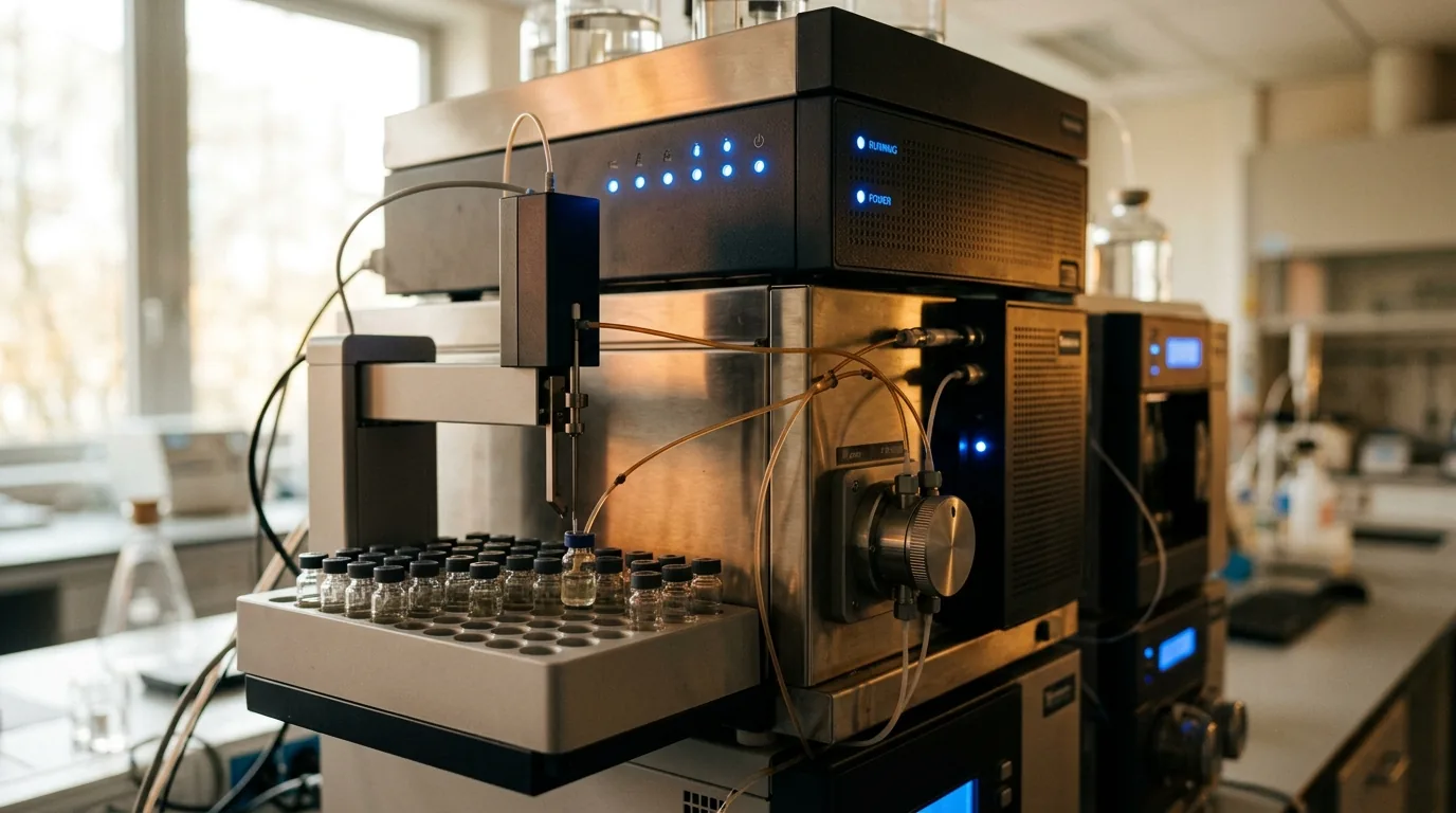
RP-HPLC on C18 columns readily separates Melanotan I and II due to their different molecular weights and hydrophobicities. Melanotan I elutes later than MT-II under standard gradients (15-60% ACN/0.1% TFA over 30 min) due to its larger hydrophobic surface area. UV detection at 220 nm (peptide bonds) and 280 nm (Trp absorbance) provides dual-wavelength confirmation [1][2].
ESI-MS confirmation: Melanotan I shows [M+2H]2+ = 824.4 and [M+H]+ = 1647.9 m/z. Melanotan II shows [M+H]+ = 1024.5 and [M+2H]2+ = 512.8 m/z. The 623 Da mass difference makes misidentification unlikely. MS/MS fragmentation of Melanotan II uniquely shows loss of the intact lactam bridge fragment, providing a definitive structural marker. Circular dichroism spectroscopy reveals distinct secondary structure profiles: Melanotan I shows a random coil/polyproline II signature, while Melanotan II displays a β-turn constrained by the cyclization [5][7].
Research Considerations and Experimental Design
When selecting between these compounds, researchers should consider their receptor selectivity requirements. Melanotan I (afamelanotide) is appropriate for MC1R-focused studies, particularly melanocyte biology and pigmentation research. Its more restricted receptor profile simplifies data interpretation when MC1R-specific effects are the research question [1][5].
Melanotan II is the compound of choice when broad melanocortin receptor activation is desired or when MC3R/MC4R signaling is the primary target. However, researchers must account for concurrent MC1R activation (pigmentation effects) and the possibility of MC5R engagement. Bremelanotide (PT-141), a metabolite of Melanotan II lacking the N-terminal acetyl-Nle, is sometimes used as an alternative with a slightly different receptor profile [2][4][6].
Side-by-Side Comparison Table
| Parameter | Melanotan I | Melanotan II |
|---|---|---|
| Full name | Afamelanotide, [Nle4,D-Phe7]-α-MSH | Ac-Nle-c[Asp-His-D-Phe-Arg-Trp-Lys]-NH2 |
| Structure | Linear, 13 residues | Cyclic lactam, 7 residues |
| Molecular weight | ~1,647 Da | ~1,024 Da |
| Key modifications | Nle4, D-Phe7 | Nle4, D-Phe7, Asp-Lys lactam |
| MC1R affinity | High (selective) | High (non-selective) |
| MC3R/MC4R affinity | Low-moderate | High |
| Pharmacophore | His-D-Phe-Arg-Trp (linear) | His-D-Phe-Arg-Trp (constrained) |
| MS [M+H]+ | 1647.9 | 1024.5 |
| Primary degradation | Deamidation, Asp isomerization | Lactam hydrolysis (ring opening) |
| Derived compounds | N/A | PT-141 (Bremelanotide) |
Frequently Asked Questions
Q: What is the key structural difference between Melanotan I and II? A: Melanotan I is a linear 13-amino-acid peptide retaining the full α-MSH backbone. Melanotan II is a truncated 7-amino-acid cyclic peptide with a lactam bridge between Asp and Lys side chains. Both share Nle4 and D-Phe7 substitutions.
Q: Which compound is more receptor-selective? A: Melanotan I shows preferential MC1R selectivity, while Melanotan II activates MC1R, MC3R, MC4R, and MC5R broadly. Neither compound has significant MC2R activity.
Q: Can they be differentiated by mass spectrometry? A: Yes, readily. Melanotan I has [M+H]+ = 1647.9 while Melanotan II has [M+H]+ = 1024.5 – a 623 Da difference. MS/MS fragmentation patterns are also completely distinct due to the linear vs cyclic architectures.
For laboratory research use only. Not for human consumption. This article presents chemical and structural data for educational and research reference purposes.
Compounds Referenced in This Article
Explore detailed chemical profiles and research guides for compounds discussed in this article:
- Melanotan 2: Complete Research Guide → /learn/melanotan-2
Further Reading on ChemVerify
- Read more: Melanotan II vs. PT-141: MSH Analog Structural Comparison → https://www.chemverify.com/learn/melanotan-2-vs-pt-141
- Read more: IGF-1 LR3 vs IGF-1 DES: Long-Acting vs Truncated Growth Factor → https://www.chemverify.com/learn/igf-1-lr3-vs-igf-1-des-comparison
- Read more: DSIP vs Selank for Sleep Research: Mechanism Comparison → https://www.chemverify.com/learn/dsip-vs-selank-sleep-research-comparison
- Read more: Follistatin vs ACE-031: Myostatin Inhibitor Comparison → https://www.chemverify.com/learn/follistatin-vs-ace-031-myostatin-comparison
References
Compare Verified Vendors
Browse COA-verified suppliers with exclusive discount codes and transparent pricing.
You Might Also Like
Continue Reading
Melanotan II vs. PT-141: MSH Analog Structural Comparison
Structural comparison of Melanotan II and PT-141 (bremelanotide) research peptides – cyclic vs linear structure, melanocortin receptor selectivity, molecular weight, and purity analysis.
IGF-1 LR3 vs IGF-1 DES: Long-Acting vs Truncated Growth Factor
Scientific comparison of IGF-1 LR3 (83aa, Arg3 substitution) and IGF-1 DES (67aa, truncated N-terminus): IGFBP binding, half-life, potency, and research applications.
DSIP vs Selank for Sleep Research: Mechanism Comparison
Scientific comparison of DSIP and Selank in sleep and anxiolytic research: delta-wave modulation, GABAergic mechanisms, peptidase resistance, and preclinical sleep architecture data.
Follistatin vs ACE-031: Myostatin Inhibitor Comparison
Scientific comparison of follistatin and ACE-031 (dalantercept) as myostatin inhibitors: binding mechanisms, activin receptor decoy strategies, preclinical muscle data, and research considerations.