
Humanin: Complete Research Guide & Chemical Profile
Complete research guide to Humanin (HN), a 24-amino-acid mitochondrial-derived peptide. Covers cytoprotective mechanisms, IGFBP-3 binding, apoptosis inhibition, and Alzheimer/diabetes research.
For laboratory research use only. Not for human consumption.
TL;DR: Humanin (HN) is a 24-amino-acid peptide encoded within the 16S ribosomal RNA gene of mitochondrial DNA. It is the founding member of the mitochondrial-derived peptide (MDP) family and exhibits potent cytoprotective activity against apoptosis, oxidative stress, and metabolic dysfunction. Key research areas include Alzheimer disease neuroprotection, IGFBP-3 binding modulation, and insulin sensitization in diabetes models.
Last verified: April 2026 | Data accuracy confirmed by ChemVerify Editorial Team
Chemical Profile & Structural Properties
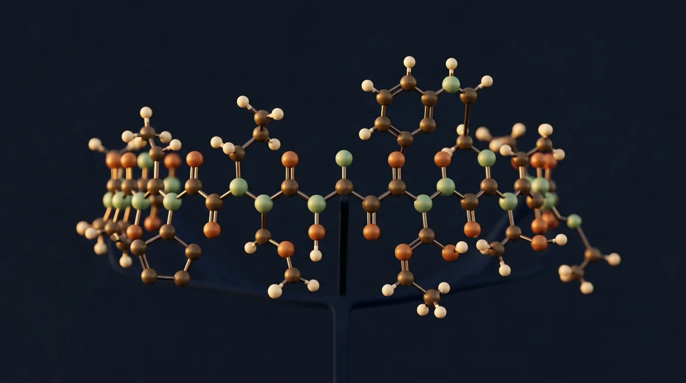
Humanin is a 24-amino-acid peptide with the sequence MAPRGFSCLLLLTSEIDLPVKRRA, a molecular weight of approximately 2,687 Da, and a molecular formula of C120H206N36O33S1. The peptide was first identified in 2001 by Hashimoto and colleagues from a cDNA library derived from the occipital cortex of an Alzheimer disease patient. Humanin is encoded within the 16S ribosomal RNA gene (MT-RNR2) of the mitochondrial genome, making it the first peptide shown to be translated from a mitochondrial open reading frame.
Structural studies using nuclear magnetic resonance (NMR) spectroscopy indicate that Humanin adopts a partially helical conformation in aqueous solution, with the central region (residues 8-19) forming an amphipathic alpha-helix. The N-terminal and C-terminal regions remain relatively disordered. The single cysteine residue at position 8 can form intermolecular disulfide bonds, allowing Humanin to dimerize. The dimeric form shows enhanced biological activity compared to the monomer in certain assay systems.
The isoelectric point (pI) of Humanin is approximately 11.2, giving it a strongly cationic character at physiological pH. This positive charge facilitates interactions with negatively charged membrane surfaces and anionic binding partners. The peptide demonstrates limited aqueous stability with a tendency toward aggregation at concentrations above 100 micromolar, necessitating careful handling in research applications including the use of fresh preparations and appropriate vehicle controls.
- Sequence: MAPRGFSCLLLLTSEIDLPVKRRA (24 amino acids)
- Molecular weight: ~2,687 Da
- Molecular formula: C120H206N36O33S1
- Gene: MT-RNR2 (mitochondrial 16S rRNA)
- pI: ~11.2 (strongly cationic)
- Structure: Amphipathic alpha-helix (central region)
- Dimerization: Via Cys8 disulfide bond
- Solubility: Aqueous; aggregation-prone above 100 µM
Mitochondrial Origin & Biosynthesis
Humanin is the founding member of the mitochondrial-derived peptide (MDP) family, which also includes MOTS-c and the small humanin-like peptides (SHLPs 1-6). These peptides are encoded within short open reading frames embedded in the mitochondrial genome, specifically within ribosomal RNA genes. The discovery that the mitochondrial genome encodes functional peptides beyond the 13 canonical oxidative phosphorylation subunits fundamentally expanded understanding of mitochondrial genetic output.
Biosynthesis of Humanin involves transcription of the MT-RNR2 gene by mitochondrial RNA polymerase, followed by translation on mitochondrial ribosomes. However, evidence also supports cytoplasmic translation of mitochondrial-derived transcripts that are exported from the organelle. Humanin is detected both intracellularly and in the extracellular space, including in plasma and cerebrospinal fluid, indicating that it functions as both an intracrine and endocrine signaling molecule. Circulating Humanin levels decline with age in both rodent models and human cohort studies.
The expression of Humanin is regulated by cellular stress conditions. Oxidative stress, endoplasmic reticulum stress, and serum starvation all upregulate Humanin expression, consistent with its proposed role as a stress-responsive cytoprotective factor. The transcriptional regulation involves retrograde signaling from mitochondria to the nucleus, linking Humanin expression to overall mitochondrial health status.
Cytoprotective Mechanisms of Action
Humanin exerts cytoprotection through multiple signaling pathways. The peptide binds to the CNTFR/WSX-1/gp130 trimeric receptor complex, activating the JAK2/STAT3 signaling cascade. STAT3 phosphorylation leads to transcriptional upregulation of anti-apoptotic genes including Bcl-2 and Mcl-1, while simultaneously suppressing pro-apoptotic BAX translocation to mitochondria. This receptor-mediated mechanism represents the primary extracellular signaling pathway for Humanin.
Intracellularly, Humanin directly binds to BAX (Bcl-2-associated X protein), preventing the conformational change required for BAX insertion into the outer mitochondrial membrane. This interaction blocks cytochrome c release and inhibits the intrinsic (mitochondrial) apoptosis pathway. The BAX-binding domain has been mapped to residues 10-19 of Humanin, overlapping with the amphipathic helical region. Additionally, Humanin interacts with tBID, another pro-apoptotic Bcl-2 family member, providing redundant protection against mitochondrial outer membrane permeabilization.
Beyond direct anti-apoptotic activity, Humanin modulates cellular stress responses through activation of the AMPK and ERK1/2 pathways. AMPK activation enhances autophagy and mitochondrial quality control through PINK1/Parkin-dependent mitophagy, while ERK signaling promotes cellular survival under oxidative stress. Humanin also reduces endoplasmic reticulum stress by modulating the unfolded protein response, specifically attenuating CHOP-mediated apoptotic signaling.
IGFBP-3 Interaction & IGF Axis Modulation
Humanin was independently identified as a binding partner of insulin-like growth factor binding protein 3 (IGFBP-3) through yeast two-hybrid screening. IGFBP-3 normally sequesters IGF-1 in the circulation, limiting its bioavailability. Humanin binding to IGFBP-3 disrupts the IGFBP-3/IGF-1 complex, potentially increasing free IGF-1 levels. This interaction has implications for GH/IGF-1 axis regulation and may partially explain the insulin-sensitizing effects observed with Humanin treatment.
The Humanin-IGFBP-3 interaction also modulates the IGF-independent pro-apoptotic activity of IGFBP-3. Nuclear IGFBP-3 promotes apoptosis through association with nur77/RXR complexes, and Humanin binding can antagonize this nuclear pro-apoptotic function. The dual modulation of both IGF-dependent and IGF-independent IGFBP-3 functions by Humanin reveals a complex regulatory node connecting mitochondrial stress signaling with the somatotropic axis.
Alzheimer Disease Research Applications
Humanin was originally discovered through its ability to protect neuronal cells from toxicity induced by familial Alzheimer disease mutant genes (APP V642I, presenilin-1, and presenilin-2 mutations). Subsequent research demonstrated that Humanin and its analogs protect against amyloid-beta peptide toxicity in primary cortical neurons and neuronal cell lines. The neuroprotective mechanism involves both the extracellular STAT3 pathway and intracellular BAX inhibition, with additional contributions from reduced oxidative stress and calcium dysregulation.
In transgenic Alzheimer mouse models (3xTg-AD, APP/PS1), systemic administration of HNG (S14G-Humanin) improved cognitive performance on Morris water maze and contextual fear conditioning tasks. Histological analysis revealed reduced amyloid plaque burden, decreased tau hyperphosphorylation, and preserved synaptic density in hippocampal and cortical regions. The mechanisms underlying amyloid reduction appear to involve enhanced microglial phagocytosis of aggregated Abeta.
Epidemiological studies have reported that circulating Humanin levels are lower in patients with Alzheimer disease compared to age-matched controls, and that Humanin levels correlate inversely with cognitive decline scores. CSF Humanin levels are being explored as a potential biomarker for mitochondrial stress in neurodegenerative disease.
Metabolic & Diabetes Research
Humanin and its analogs demonstrate significant insulin-sensitizing activity in preclinical models. In high-fat diet-fed mice and genetically obese (ob/ob) models, HNG administration improved glucose tolerance, reduced hepatic glucose output, and decreased circulating insulin levels indicative of improved insulin sensitivity. The metabolic effects are mediated through hypothalamic signaling (central STAT3 activation) and direct peripheral actions on skeletal muscle and adipose tissue.
At the cellular level, Humanin enhances insulin signaling by promoting IRS-1 tyrosine phosphorylation and downstream Akt activation. Humanin also reduces lipotoxicity in pancreatic beta-cells by attenuating palmitate-induced ER stress and apoptosis, suggesting a protective role for islet preservation. In liver cells, Humanin suppresses gluconeogenic gene expression (PEPCK, G6Pase) through STAT3-mediated transcriptional repression.
The age-dependent decline in circulating Humanin has been proposed as a contributing factor to age-related metabolic deterioration. Cross-sectional studies demonstrate that plasma Humanin levels correlate negatively with HOMA-IR (insulin resistance index) and positively with insulin sensitivity measures. These associations remain significant after adjustment for age, BMI, and other metabolic covariates.
Cardiovascular Research Applications
Humanin shows cardioprotective properties in ischemia-reperfusion injury models. In isolated heart preparations and in vivo coronary artery occlusion models, Humanin pretreatment reduced infarct size, improved contractile recovery, and decreased cardiomyocyte apoptosis. The cardioprotective mechanism involves preservation of mitochondrial membrane potential, reduced cytochrome c release, and activation of the RISK (Reperfusion Injury Salvage Kinase) pathway including Akt and ERK1/2.
In atherosclerosis models, Humanin reduces endothelial dysfunction by suppressing oxidative stress and inflammatory cytokine expression. ApoE-knockout mice treated with HNG showed reduced atherosclerotic plaque area and decreased macrophage infiltration. Humanin also attenuates angiotensin II-induced cardiac hypertrophy through suppression of ROS production and calcineurin/NFAT signaling.
Humanin Analogs & Structural Modifications
Structure-activity relationship studies have identified several key residues critical for Humanin biological activity. Alanine-scanning mutagenesis revealed that Pro3, Ser7, Cys8, Leu9, Leu12, Thr13, Ser14, and Pro19 are essential for cytoprotective function. The most important substitution is S14G (serine-14 to glycine), generating HNG with approximately 1,000-fold greater potency than native Humanin.
Additional analogs include HNG-F6A (degradation-resistant), C-terminal amidation variants (improved stability), and Colivelin, a hybrid peptide combining modified Humanin with the ADNF fragment for dual-mechanism neuroprotection. D-amino acid substitutions provide enhanced protease resistance for in vivo studies.
- HNG (S14G-Humanin): ~1,000x more potent than native HN
- HNG-F6A: Degradation-resistant analog
- Colivelin: AGA-HNG fused with ADNF fragment
- C-terminal amidation: Improved metabolic stability
- D-amino acid substitutions: Enhanced protease resistance
- Rat/mouse Humanin: Identical sequence (highly conserved)
Analytical Methods & Detection
Quantification of Humanin in biological samples employs several complementary analytical approaches. ELISA using anti-Humanin antibodies provides detection limits in the low picomolar range. However, cross-reactivity with endogenous Humanin-like peptides (SHLPs) must be considered when interpreting results from biological matrices.
LC-MS/MS with stable isotope-labeled internal standards enables absolute quantification with minimal cross-reactivity. Typical plasma concentrations in healthy adults range from 0.5-2.0 ng/mL. For synthetic preparations, MALDI-TOF-MS identity testing and RP-HPLC purity assessment (>95% specification) represent standard quality control measures.
References & Further Reading
The following publications represent foundational and recent research on Humanin and mitochondrial-derived peptides.
Further Reading on ChemVerify
- Read more: TRH (Thyrotropin-Releasing Hormone): Research Guide & Chemical Profile → https://www.chemverify.com/learn/trh-thyrotropin-releasing-hormone-research-guide
- Read more: Ipamorelin + CJC-1295 (No DAC) Stack: Synergy Research Guide → https://www.chemverify.com/learn/ipamorelin-cjc-1295-no-dac-stack-synergy
- Read more: TP508 (Chrysalin): Research Guide & Chemical Profile → https://www.chemverify.com/learn/tp508-chrysalin-research-guide-chemical-profile
- Read more: Semax for Cognitive Research: ACTH(4-10) Analog Mechanism → https://www.chemverify.com/learn/semax-cognitive-research-acth-mechanism
References
Compare Verified Vendors
Browse COA-verified suppliers with exclusive discount codes and transparent pricing.
You Might Also Like
Continue Reading
Ipamorelin + CJC-1295 (No DAC) Stack: Synergy Research Guide
Research guide to the Ipamorelin + CJC-1295 No DAC growth hormone stack: GHRH-GHRP synergy mechanism, GH pulse amplification, timing protocols, and reconstitution guidance.
Semax for Cognitive Research: ACTH(4-10) Analog Mechanism
Semax mechanism of action: ACTH(4-10) fragment with Pro-Gly-Pro modification, BDNF and NGF upregulation, BBB penetration, nasal delivery, and Russian clinical research history.
TRH (Thyrotropin-Releasing Hormone): Research Guide & Chemical Profile
TRH (pGlu-His-Pro-NH2) is a hypothalamic tripeptide releasing TSH and prolactin. MW 362.38, neuroprotective research, and neuroendocrine signaling reviewed.
TP508 (Chrysalin): Research Guide & Chemical Profile
TP508 (Chrysalin) is a 23-amino-acid thrombin receptor binding domain peptide with FDA orphan drug status for fracture healing. Bone repair and angiogenesis research.